How to Know If Your Growth Plates Are Closed: 4 Methods Ranked by Accuracy
Whether you are a teen tracking your growth, a parent checking on your child, or an athlete wondering if your bones are still developing — this guide covers every method available, what each one can actually tell you, and when you need a doctor involved.
The only definitive way to know if growth plates are closed is a bone age X-ray of the left hand and wrist, interpreted by a doctor. At-home methods — tracking height changes, age, and puberty stage — can give you a strong probability estimate, but not a confirmed answer. This guide explains all four methods, what they show, and their limits.
What Growth Plates Actually Are (And Why Closing Matters)
Growth plates — known medically as epiphyseal plates or physes — are thin zones of cartilage sitting at each end of the body’s long bones: the femur, tibia, fibula, radius, ulna, humerus, and the small bones in your hands and feet. Their job is simple and critical: they manufacture new bone tissue, pushing bones longer and making you taller.
While the plates are active, this cartilage is softer than the surrounding bone — which is why growth plate fractures in children are so clinically significant. A hard impact hits the weakest point first, and in a growing child, that is often the growth plate, not the bone shaft. According to NYU Langone Health, up to 30% of fractures in children can occur around growth plates.
As puberty progresses, rising levels of estrogen — in both females and males — signal the chondrocytes (cartilage cells) in the growth plate to stop dividing. Over time the cartilage thins, bridges form across it, and eventually it is completely replaced by solid bone. That moment is called epiphyseal closure, or growth plate fusion. Once it happens in a given bone, that bone cannot grow longer. The window is gone.
Not all growth plates close at the same time. The hand and foot plates can be fully fused while the knee and hip plates are still open and contributing to height. This means partial closure is common — and a closed wrist X-ray does not always mean you have stopped growing entirely.
4 Methods to Check If Growth Plates Are Closed — Ranked by Accuracy
Not all methods are equal. Here they are in order of reliability, from most accurate to least:
The left hand is used for bone age studies because most people are right-hand dominant. The dominant hand is more likely to have sustained minor injuries that could affect growth plate appearance. Using the non-dominant hand gives a more representative, undisturbed image. Orthopedic surgeons also use specific X-ray markers from the pelvis (the Risser sign) for a secondary estimate of skeletal maturity, especially in scoliosis management.
Age Ranges by Sex: When Growth Plates Typically Close
Timing varies significantly between individuals — and between bones within the same person. That said, sex-based averages provide a useful frame of reference. According to Duke Health pediatric orthopedics, most children grow an average of two additional years after completing their most rapid pubertal growth spurt.
| Bone / Location | Girls (approx.) | Boys (approx.) | Notes |
|---|---|---|---|
| Fingers and hand bones | 12–14 years | 14–16 years | Among the earliest to close; hands reach adult size before height |
| Elbow | 12–14 years | 14–16 years | Closes relatively early in the sequence |
| Wrist (distal radius) | 14–16 years | 16–18 years | Standard site used for bone age X-ray assessment |
| Ankle | 14–16 years | 16–17 years | Foot and ankle close before knee in most cases |
| Knee (distal femur/proximal tibia) | 15–17 years | 17–19 years | One of the last major height-contributing plates to close |
| Hip and pelvis | 15–20 years | 17–21 years | Iliac crest can remain partially open into early adulthood |
| Shoulder and clavicle | 17–20 years | 18–25 years | Among the last plates to fully fuse in the skeleton |
These ranges reflect population averages and have meaningful variation. According to documented skeletal development research, complete fusion can occur as early as age 12 in girls (though 14–15 is most common) and as early as 14 in boys (with 15–17 being most typical). The clavicle and medial end of the collarbone are often the last bones in the entire skeleton to fully fuse, sometimes not completing until the mid-20s.
Which Growth Plates Close First — and Which Close Last
Closure follows a broadly predictable sequence across the skeleton, generally moving from the extremities (hands, feet) toward the central body (spine, pelvis). Understanding this sequence explains why a child whose hand plates have closed may still gain height from plates in their legs.
-
1stFingers, toes, and small foot bonesThe phalanges and smaller foot bones are typically the earliest to fuse. Hands and feet reach their adult proportions well before the body’s overall height is set.
-
2ndElbow and ankle regionThe elbow joint plates close earlier than the knee plates. Ankle plates follow a similar early pattern. Neither area contributes much to overall standing height.
-
3rdWrist (distal radius and ulna)The wrist is the clinical reference point for bone age X-rays. Closure here is a strong indicator of approaching skeletal maturity, but does not guarantee the leg plates are done.
-
4thKnee (distal femur, proximal tibia)The knee’s plates are among the most important for height because the femur (thigh bone) is the longest bone in the body. Closure here signals that the majority of height gain is complete. Research published in PMC confirms females complete this stage 1–2 years earlier than males.
-
LastShoulder, clavicle, and iliac crest (pelvis)The medial clavicle and iliac crest of the pelvis are typically the final plates to fuse in the human skeleton, sometimes completing in the early-to-mid 20s. These closures contribute little to height but are used in forensic age estimation and medical contexts like scoliosis surgery timing.
What Speeds Up or Delays Growth Plate Closure
Several factors influence when growth plates close — some within your control, most not. Understanding them explains why two people of the same age can be at very different stages of skeletal maturity. Research reviewed in PMC’s puberty and epiphyseal fusion review confirms that hormonal signaling — particularly estrogen — is the dominant driver of closure timing.
Estrogen levels — the primary hormonal trigger for closure in both sexes. Higher estrogen accelerates the timeline. Girls close earlier partly because they reach higher estrogen levels sooner.
Genetics — the single strongest predictor of when your plates close. Children tend to close within a narrow window of their parents’ skeletal maturity patterns.
Precocious puberty — early puberty means earlier estrogen spike, which can close plates prematurely and result in shorter adult stature than genetic potential would predict.
Nutrition — chronic malnutrition or deficiencies in calcium, vitamin D, and protein can delay skeletal maturation. Adequate nutrition supports plates reaching their full potential but does not keep them open longer.
Chronic illness — conditions affecting hormones or bone metabolism (thyroid disorders, growth hormone deficiency, kidney disease) can significantly alter closure timing.
Growth plate injury — direct trauma to a growth plate can cause early or abnormal closure in that specific location, potentially leading to uneven limb length or bone curvature.
Ethnicity — documented variation exists in bone age across different ethnic populations, which is why some bone age atlases have been developed using ethnically diverse reference groups.
Body weight/BMI — higher BMI in adolescents has been associated with earlier skeletal maturation in some studies, possibly through higher circulating estrogen from adipose tissue.
Normal physical activity and exercise do not accelerate growth plate closure. A cross-sectional MRI study of 958 adolescents found no significant correlation between physical activity levels and timing of growth plate fusion. Everyday sports, running, and gym use are safe. The risk comes specifically from direct growth plate injury — trauma, not regular exercise.
Physical Signs That Growth Has Probably Stopped
If you cannot or do not want to get an X-ray, these observable signs can give you a reasonable probability estimate. None of them are confirmations — but together, they build a picture.
No Height Change for 12+ Months
The most practical at-home indicator. Measure at the same time of day (morning is most accurate — you are slightly taller after sleeping), against the same wall, using consistent technique. If three or more measurements over 12 months show no change, the plates contributing to height are likely closed or very close to it.
Puberty Is Complete
Growth plates respond to the hormonal environment of puberty. Once puberty is fully complete — including cessation of voice changes, body hair progression, and other secondary sex characteristics — the hormonal signals that drive bone elongation have diminished. The Nemours KidsHealth bone age guide describes how doctors use puberty staging alongside X-rays to interpret skeletal maturity.
You Are Within Expected Range of Parental Height
A pediatric formula called mid-parental height helps predict a child’s genetic height ceiling. If you have essentially reached this predicted height and puberty is complete, continued growth is unlikely. Ask your pediatrician to calculate mid-parental height if you have not had it done.
Your Shoe Size Has Not Changed in Over a Year
Foot bones (smaller than long bones) close earlier. Stable shoe size for over a year suggests the extremities are done. This is not reliable for overall height prediction — you can stop growing in your feet while the knee plates are still active — but it supports the overall picture when combined with the other indicators above.
You cannot feel your growth plates from outside the body. Cartilage sits deep within joint structures, well beneath skin, muscle, and other tissue. Any claim that you can “feel a gap” or assess closure by touching your joints is not medically valid. The plates are not surface-accessible without imaging.
What the Bone Age X-Ray Actually Shows
A bone age study uses a single X-ray of the left hand and wrist. The resulting image is compared against a standardized reference atlas — the most widely used being the Greulich and Pyle atlas, developed from a large sample of children across different developmental stages.
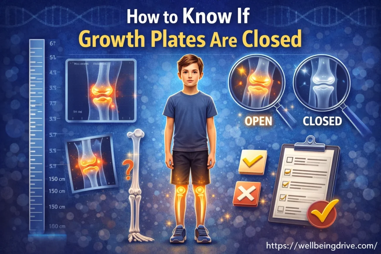
On the image, growth plates that are still open appear as dark lines or gaps at the ends of bones. These gaps represent cartilage, which does not absorb X-ray radiation as strongly as bone and therefore appears darker. When a plate closes, those gaps fill in with solid bone and disappear from the image.
A radiologist or pediatric orthopedic surgeon assigns a “bone age” — a skeletal maturity estimate — by comparing the image to the atlas. A child whose bone age matches their chronological age is developing on a typical schedule. A bone age that is significantly older or younger than the child’s actual age can signal early or delayed development that may warrant investigation.
When X-ray results are ambiguous — or when a doctor needs to assess partial closure or growth plate injury in detail — MRI is the preferred follow-up. Unlike X-rays, MRI can visualize cartilage directly, including the individual sublayers of the growth plate (resting zone, proliferative zone, hypertrophic zone). It can identify areas of partial bridging that are not visible on a standard radiograph.
When to Actually See a Doctor About Growth Plates
Curiosity about whether you have stopped growing is normal and does not require a doctor’s visit. However, there are specific situations where professional assessment genuinely matters — for clinical reasons, not just peace of mind.
- A child is significantly shorter than expected based on parental heights and growth charts, and no obvious cause has been identified
- A child appears to have stopped growing unusually early (before age 12 for girls, before age 14 for boys)
- A teenager is still growing well past the typical age cutoff — especially if they are experiencing bone or joint pain alongside it
- There has been a direct injury to a joint area in a child or adolescent, with persistent pain or visible deformity — growth plate fractures need prompt attention to prevent long-term complications
- A child has been diagnosed with a condition affecting hormones, nutrition, or kidney function, and you want to monitor bone development as part of management
- An athlete (especially in high-impact or contact sports) has recurring pain at a joint that does not resolve — overuse growth plate injuries are common in young athletes
- There is a suspected leg length discrepancy or bone curvature developing in a still-growing child
For routine growth curiosity, a simple conversation with a pediatrician and a standard growth chart review is usually enough. An X-ray is not needed just to satisfy curiosity about whether you are still growing — but if it matters for a clinical decision, it is a fast, low-radiation, inexpensive test that gives a direct answer.
FAQs
You cannot know for certain without medical imaging, but strong indicators include no measurable height increase in 12 or more months, full completion of puberty (including cessation of other physical changes), and being past the typical age range for your sex (girls: 14–16, boys: 16–18). These are probability estimates, not proof. A left-hand X-ray is the standard method to confirm closure.
For girls, most growth plates close between ages 13 and 15, with full skeletal maturity typically by 16–17. For boys, closure generally occurs between 15 and 17, with the last plates (shoulder and clavicle) closing as late as the early to mid-20s. Age varies significantly based on genetics, hormones, and ethnicity. No single age applies to everyone.
The last growth plates to close are typically around the shoulder, clavicle, and pelvis (iliac crest). The medial clavicle may not fully fuse until the mid-20s. The knee plates — distal femur and proximal tibia — are among the last of the major height-contributing plates to close, which is why they are clinically important for height prediction. Hands and feet close first.
Yes. An X-ray of the left hand and wrist is the standard method for assessing skeletal maturity. Open growth plates appear as dark lines (gaps of cartilage) at the ends of bones. Closed plates look solid and fused, with no visible gap. A pediatric radiologist or orthopedic specialist interprets the results and assigns a bone age estimate based on a standardized atlas comparison.
Yes. Premature closure can result from precocious (early) puberty, abnormally high estrogen levels, certain growth hormone conditions, direct injury to the growth plate, radiation exposure, or infection. Early closure means the bone stops growing before reaching its genetic potential, which can result in shorter stature or, if closure happens unevenly across the plate, a curved or deformed bone.
Normal physical activity does not accelerate growth plate closure. A cross-sectional MRI study of 958 adolescents found no significant correlation between physical activity and the timing of fusion. However, direct trauma to a growth plate from injury or overuse in high-impact sports can cause premature or abnormal closure in that specific location — which is why growth plate injuries in young athletes need prompt medical attention.
Medical Disclaimer: This article is written for informational and educational purposes only and does not constitute medical advice, diagnosis, or treatment. Information on growth plate closure ages, imaging methods, and developmental timelines is sourced from peer-reviewed literature, Duke Health, NYU Langone Health, Nemours KidsHealth, and published research available via PubMed and PMC. Individual bone development varies. Always consult a licensed pediatrician, orthopedic surgeon, or healthcare provider for assessment of specific growth and development concerns.
Disclaimer: The content on Wellbeingdrive is for informational purposes only and not a substitute for professional advice. Always consult a qualified expert for health concerns.